Ready to get on top of this?
📞 Call Now — speak with our team
🗓 Book Online — available 24/7
What Is the Anterior Oblique Sling?
The anterior oblique sling is a diagonal myofascial chain that connects the trunk to the contralateral lower limb across the anterior pelvis and abdominal wall. It includes:
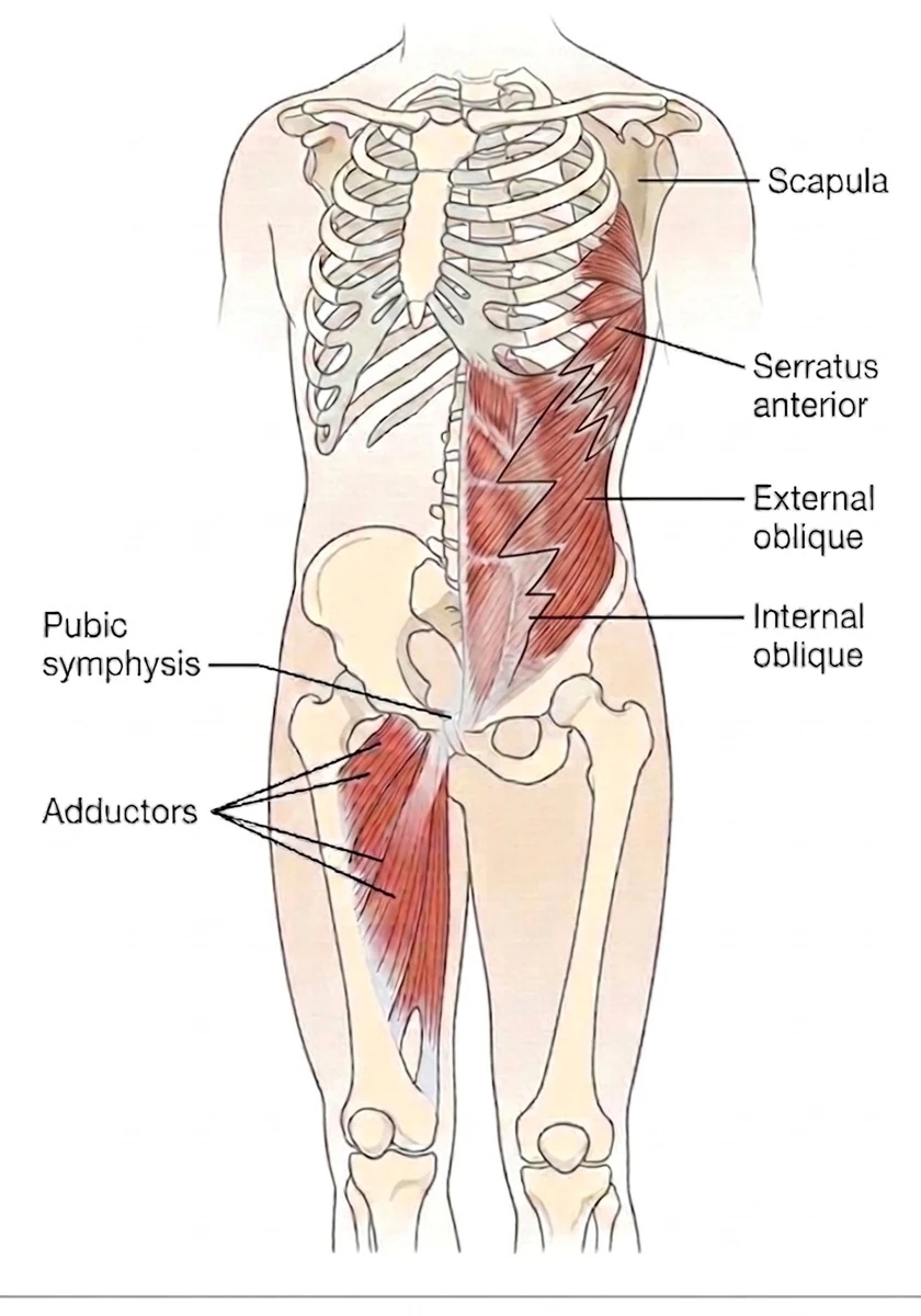
- Internal oblique and transversus abdominis — the deep anterolateral abdominal muscles, generating tensile force through the anterior abdominal fascia
- Anterior abdominal fascia (anterior rectus sheath and linea alba) — the connective tissue bridge across the midline through which diagonal tension is transmitted
- Contralateral external oblique — completing the diagonal on the opposite side of the anterior trunk
- Contralateral adductor muscles (particularly adductor longus) — the lower limb anchor, connected through the inguinal and pubic fascial complex
- Serratus anterior — the upper anchor. The external oblique is fascially continuous with the serratus anterior above it, which carries the chain onto the scapula
The popular description of what crosses the midline here is wrong. There is no thick sheet of tissue joining the abdominal muscles straight onto the adductor: the connection is thin, and the rectus abdominis does not reach in front of the pubic bone at all. Two separate dissection studies agree on that much.
They also reported something more striking: that adductor longus tendon fibres insert perpendicularly into bone through a fibrocartilage enthesis and cross-connect along the anterior pubic ligament into the tendon on the opposite side, which they proposed as a reason groin symptoms are so often bilateral. That particular finding is disputed. A 2024 dissection study describes the region differently, and we have set out the disagreement in the research section below rather than pick a side here. What is not in dispute is that something connects the two halves of the pelvis at the pubis — and that load applied to one side is shared by both.
In our clinical experience, that observation seems to extend further than the groin. What often surprises patients is that we sometimes find the restriction on the opposite side to the symptoms — a densified ankle or foot on one side alongside hip pain on the other, for instance. We want to be clear that this is a clinical observation: no study we are aware of has tested it. We mention it because it is part of why we assess both sides rather than only the painful one.
The thoracolumbar fascia also contributes to the posterior anchor of the abdominal muscles through the lateral raphe — connecting the anterior diagonal tension of the AOS to the paraspinal system (Schuenke et al., 2012; Vleeming et al., 2014). The sling is therefore not isolated to the anterior trunk: it is mechanically integrated with the broader lumbopelvic fascial system.
The Lower End: Shared Tissue at the Pubis
At the pubis the abdominal and adductor tissues do not simply meet end to end. Dissection shows the external oblique aponeurosis running directly into the adductor longus aponeurosis as one continuous plate, with a second plate linking the rectus abdominis and pyramidalis to the gracilis and adductor brevis.
This is not a tendon handing off to another tendon. It is shared tissue, which is why the region tends to fail as a unit rather than as one structure — see groin pain for what that looks like clinically.
The Upper End: Serratus Anterior and the Shoulder
The anterior oblique sling is usually described from the adductors up to the opposite side of the abdomen, and the description typically ends there. The full sling runs from hip to shoulder across the trunk. The external oblique does not end at the ribs — its fascia is continuous with that of serratus anterior, the muscle that holds your shoulder blade against the ribcage and rotates it upward as you lift your arm.
The clinical consequence is that a diagonal problem in the trunk can present at the shoulder rather than the groin. If serratus anterior is not doing its job, the scapula cannot rotate upward properly, and the mechanical result is the same as the more familiar scapular causes: less room beneath the acromion during elevation. This is one of the reasons we assess the trunk and the opposite hip in people who present with shoulder impingement or rotator cuff pain, rather than assessing the shoulder alone.
That is not only an anatomical inference. When serratus anterior activity was measured by EMG in twenty-one healthy adults, loading the opposite leg and the trunk significantly increased serratus activation compared with the same arm movement performed on its own — including in both of the conditions designed specifically to load the diagonal (Kaur et al., 2014). That higher activation might mean the scapula simply needs more stability against a larger load arriving from the legs; it might reflect neural coordination across the chain; or it might be force transfer along the sling itself. The effect was clear and consistent. The study was not built to separate those three, and cannot tell us which was at work.
Beyond the scapula the chain does not simply stop: the fascia enveloping serratus anterior continues posteriorly with that of the rhomboids at the medial border of the scapula — Stecco describes this as the serratorhomboid complex — which is where this anterior chain meets the posterior structures of the shoulder girdle. That is a longer story than this page needs, but it is worth knowing the chain keeps going.
What Does It Do?
Anterior Trunk Stabilisation During Rotation
The anterior oblique sling is the primary anterior stabiliser of the trunk in the transverse plane. During any rotational movement — walking, running, throwing, swinging, kicking — the diagonal tension of the AOS resists excessive trunk rotation and manages the transfer of force from the lower limb into the trunk and vice versa.
As the right leg swings forward in gait, the right hip flexes and the trunk begins to counter-rotate to the right. The left internal oblique and the right external oblique (which share fascial continuity across the anterior abdominal wall) generate a diagonal tensile force that manages this rotation and couples the swing leg to the contralateral trunk. The adductor longus on the right side, pulling the femur toward the midline, is simultaneously loaded — and its fascial continuity with the contralateral (left) abdominal aponeurosis at the pubic symphysis means that this adductor loading is transmitted diagonally through the anterior pelvis into the opposite trunk.
Hip Adductor and Pubic Symphysis Load Management
The pubic symphysis is the mechanical hub of the anterior oblique sling. Forces generated by the trunk muscles on one side and the hip adductors on the other converge at this point. In a well-functioning AOS, these diagonal tensions are balanced and distributed — the pubic symphysis experiences controlled, symmetric compressive loading from both sides of the chain.
When the sling is restricted or asymmetric — through a unilateral abdominal restriction, an adductor strain, or a fascial densification at the inguinal or pubic region — the force distribution at the pubic symphysis becomes asymmetric. If one side of the symphysis then receives more shear than the other, this can contribute to the pattern of groin pain, lower abdominal pain, and adductor loading that characterises pubic symphysis-related dysfunction and athletic groin presentations.
Integration with the Thoracolumbar Fascia
The abdominal muscles — particularly the transversus abdominis and internal oblique — insert posteriorly into the thoracolumbar fascia via the lateral raphe (Schuenke et al., 2012). This means the anterior oblique sling is not mechanically independent of the posterior system. When the AOS generates anterior diagonal tension, it also places tensile load on the TLF posterior layer through these posterior abdominal attachments. The result is a coordinated circumferential tensioning of the lumbopelvic fascial envelope — the anterior and posterior slings working in functional partnership to provide three-dimensional load sharing.
Vleeming et al. (2014) described this coupling between the deep abdominal muscles and the paraspinal system through the TLF, demonstrating that the abdominal and posterior trunk systems are not separate stabilisers but components of an integrated force-transfer mechanism.
When It Goes Wrong: Clinical Relevance
Adductor-Related Groin Pain
Groin pain in athletes is one of the most common and diagnostically complex presentations in sport. The adductor muscles, the inguinal region, the pubic symphysis, and the hip itself can each contribute — and combinations of contributing structures are common. The anterior oblique sling framework explains why: the adductor longus, the abdominal muscles, and the pubic symphysis are not isolated structures but components of a diagonal chain, and dysfunction anywhere in the chain alters the loading at every other point.
Ahmadi et al. (2025) demonstrated that an exercise programme specifically targeting the anterior oblique sling produced significant reductions in groin pain in soccer players compared to a standard strengthening programme — providing clinical evidence that addressing the chain as a whole, rather than the isolated adductors or abdominals, may support more complete recovery from athletic groin pain.
The Asymmetric Load Pattern
Because the sling is a diagonal, a restriction on one side does not stay on that side. It changes how load is shared across the pubic symphysis, and the symptoms may appear opposite the restriction rather than over it. This is a pattern that is frequently not identified where the inguinal or adductor region is assessed in isolation, without reference to the broader diagonal.
The Kicking Athlete
In sports involving repeated unilateral kicking — football (Australian rules, soccer, rugby league), martial arts — one diagonal of the AOS is loaded substantially more heavily than the other. The dominant-side adductor loading generates repeated diagonal tension through the pubic symphysis into the contralateral abdominal wall with each kick. Over time, this may produce asymmetric fascial stress at the pubic symphysis and the inguinal region — a pattern that is directly relevant to the high prevalence of groin pain and pubic symphysis dysfunction in kicking sports.
The Desk Worker and Anterior Abdominal Restriction
Prolonged sitting tends to reduce available hip extension and, separately, progressively inhibits the deep abdominal muscles. These travel together because they share a cause, not because one produces the other. When the internal oblique and transversus abdominis are inhibited, the anterior oblique sling is unable to generate its normal diagonal tension — the abdominal anchor of the chain is no longer contributing, and the adductor longus and inguinal region must absorb more of the functional load without the benefit of the diagonal chain's distributed force sharing.
This is a common contributing factor in groin and adductor presentations in desk workers who also train — the AOS is already in a compromised state before the training load is applied.
Anterior Pelvic Floor and Lower Abdominal Contributions
The pelvic floor muscles share anatomical territory with the inferior margin of the anterior oblique sling — the internal obturator, the pubococcygeus, and the deep transverse perineal muscles all converge in the vicinity of the pubic symphysis and the inguinal region. Restriction in the pelvic floor fascial environment can alter the tension distribution at the inferior anchor of the AOS, contributing to lower abdominal and inguinal symptoms that are not explained by isolated abdominal or adductor assessment.
The Fascial Lens: Why We See This Differently
For the anterior oblique sling to function normally, the fascial structures that connect its components must be working normally too — the anterior abdominal aponeurosis, the inguinal ligament and inguinal ring fascial complex, and the pubic fascial attachment of the adductor longus tendon. Dissection work shows these are not a series of separate attachments that happen to sit near one another: they are connected tissue, arranged in layers, with continuity across the midline at the pubis. Exactly which structures form that continuity is disputed, and we set it out in the research section below.
When any section of this chain becomes densified — losing its normal inter-layer gliding capacity — the coordination across the diagonal can be disrupted. The abdominal muscles can still contract, the adductors can still fire, but the timing and sharing of load between them is altered. Normal load sharing across the tissues that make up the sling is compromised, and the site where that altered load is felt (as pain, or as restriction) varies with the movement and loading demands of the individual.
This is clinically important for two reasons:
The restriction may be remote from the pain. A fascial restriction in the right anterior abdominal wall — perhaps from an old abdominal strain, a previous appendectomy scar, or sustained anterior pelvic tilt loading — may alter the force distribution in the right-to-left diagonal without producing right-sided symptoms. The left-sided groin or adductor loading increases, and left-sided symptoms develop — even though the relevant restriction is on the right. Assessment of the full diagonal, not only the symptomatic side, is required to identify this pattern.
Strengthening alone is insufficient when the fascial environment is restricted. An adductor strengthening programme addresses the muscular anchor of the anterior oblique sling. But if the fascial environment between the adductor longus, the inguinal region, and the anterior abdominal aponeurosis is restricted and poorly gliding, strengthening the muscle does not restore the smooth diagonal force transmission the chain requires. The strength is there; the fascial connectivity is not.
The Fascial Picture — Anterior Oblique Sling
The anterior oblique sling works as a diagonal across the front of the trunk, linking the abdominal muscles on one side to the hip adductors on the other through the fascial tissues that meet at the pubic symphysis. Its effectiveness depends not only on the strength of its muscular components, but on the gliding capacity of the fascial structures that connect them across the inguinal and pubic region. Densification anywhere in this chain — in the anterior abdominal fascia, the inguinal region, or the adductor-pubic junction — can disrupt how that diagonal coordinates, and the load it would otherwise have shared is taken up somewhere else along it. That is one reason the painful site and the restricted site are rarely the same place. A treatment approach that addresses the fascial environment of the full chain, not only its individual muscular components, is directed at the mechanism underlying these presentations.
What Does the Research Say?
The Anatomy at the Pubis Is Genuinely Contested
The anterior oblique sling is a functional model. It rests on the continuity of the anterior abdominal fascia across the midline, on the coordinated timing of the muscles involved, and on force-closure mechanics at the pelvis — not on a single uninterrupted strand of tissue running from one oblique into the opposite adductor. That distinction matters, because the fine anatomy at the pubic symphysis is one of the more actively disputed areas in musculoskeletal research.
Several groups have looked at the same few square centimetres of tissue and described it differently. The disagreement is real, and it is worth knowing about if you have been given a confident anatomical explanation for your groin pain.
The three dissection studies, and where they disagree
- Schilders and colleagues (2017) described the pyramidalis–anterior pubic ligament–adductor longus complex, or PLAC, and found that the rectus abdominis is not attached to the adductor longus.
- De Maeseneer and colleagues (2019) agreed there is no thick aponeurotic plate, measuring the abdominal-to-adductor connection at roughly 1.5 mm, and reported that adductor longus tendons cross-connect through the anterior pubic ligament into the tendon on the opposite side.
- Tharnmanularp and colleagues (2024) dissected fourteen pelvic halves and described two distinct layers instead: a superficial external oblique–adductor longus aponeurosis on the same side, and a deeper rectus abdominis–pyramidalis–gracilis–adductor brevis aponeurosis that does fuse across the midline. On their account the adductor longus is not part of the structure that crosses, and they state that their findings oppose both of the models above.
They agree on more than the disagreement suggests. The old idea of a thick aponeurotic plate joining rectus abdominis to adductor longus is now rejected by all of them. And something does cross the midline at the pubis. What remains unsettled is which structures form it.
Some of that variation is probably real biological variability. Some of it is methodological: small samples, different thresholds for what counts as tissue fusion rather than simple apposition, and cadaveric material that is typically elderly — the donors in the 2024 study averaged 81 years — being used to reason about injuries that occur mostly in young athletes.
Our position is that the sling is best understood as a functional and clinical model, supported by coordination and outcome evidence, and that the fine anatomy at the pubis is an open question. Force does not require one continuous strand of collagen to transfer between structures; shared fascial sheets, adjacent connections and a common bony attachment will do it. The clinical case for assessing the chain rather than the sore part does not stand or fall on which dissection study turns out to be right.
Clinical Evidence in Athletic Groin Pain
Ahmadi et al. (2025) conducted a randomised controlled trial in soccer players with chronic groin pain, comparing an anterior oblique sling-specific rehabilitation programme to a standard adductor and abdominal strengthening protocol. The AOS-specific programme produced significantly greater reductions in pain and improvements in functional performance — suggesting that addressing the diagonal chain as a whole, including its fascial connectivity, produces better clinical outcomes than isolated local strengthening.
Abdominal-Paraspinal Coupling Through the TLF
Vleeming et al. (2014) described the mechanical coupling between the deep abdominal muscles and the paraspinal system through the thoracolumbar fascia — demonstrating that the abdominal and posterior trunk systems are integrated force-transfer mechanisms rather than independent stabilisers. This work contextualises the AOS within the broader lumbopelvic fascial system and explains why anterior chain restriction can contribute to posterior trunk pain, and vice versa.
The Lateral Raphe as Anterior-Posterior Integration Point
Schuenke et al. (2012) characterised the lateral raphe and the thoracolumbar composite at the lumbosacral base — the anatomical points at which the abdominal aponeuroses connect with the posterior paraspinal system. This work establishes the structural basis for the integration between the anterior oblique sling and the TLF, explaining how anterior abdominal restriction can alter posterior trunk mechanics and contribute to lumbosacral loading.
Does Force Actually Cross the Midline?
The anatomy is consistent. Norton-Old et al. (2013) dissected the region and found the adductor longus tendon communicated with the contralateral rectus sheath across the symphysis in most of the specimens they examined. Whether meaningful force actually crosses that junction has been directly tested once — in embalmed cadavers, under a passive applied load — and was not demonstrated. We would not expect it to have been.
Force transmission through fascia depends on the position the joints are held in (Mohr et al., 2023), on the tissue behaving normally (Procópio et al., 2025), and on it being loaded actively rather than pulled on (Carvalhais et al., 2013). Preserved cadaveric tissue under a passive load is the least likely place to detect it. The same authors call for the work to be repeated in unembalmed tissue. When the equivalent chain at the back of the body was tested in living people with active muscle contraction, the effect was clear — and when that same test was run in people with chronic low back pain, the local response survived while the distant one disappeared.
So a null result in preserved tissue tells us more about the specimen than about the pathway. That the phenomenon is real is not in doubt: Ajimsha et al. (2022), in a scoping review of in-vivo evidence, found myofascial force transfer between anatomically connected muscles demonstrable in living subjects across several regions of the body. What is missing is evidence for this particular diagonal. Krause et al. (2016), reviewing the measured force-transfer studies, found the front functional line rested on a single study showing a slight transfer that did not reach statistical significance; and Kretschmer and Wilke (2026), in the first meta-analysis of remote fascial stiffness, note that of fifteen studies none examined the anterior diagonal at all.
We therefore treat this chain as an anatomical and coordination relationship, which the evidence supports, and regard the force-transmission question as open rather than answered.
How We Assess and Address This
Our assessment of the anterior oblique sling evaluates the full diagonal chain across both sides:
- Adductor longus length and load tolerance — passive and active adductor assessment, including adductor squeeze testing and loaded hip adduction under resistance; noting asymmetry between sides
- Inguinal and pubic region palpation — direct assessment of the inguinal canal, inguinal ring, and pubic aponeurotic attachment, identifying fascial restriction and tenderness that may indicate densification at the sling's midline hub
- TLF and abdominal fascia assessment using Stecco FM protocols — palpatory verification of the anterior chain CCs from the anterior abdominal wall to the inguinal and pubic region; identifying the most densified points in the diagonal
- Hip and pelvic movement pattern assessment — observing rotational loading tasks (single-leg stance, rotational activities) to identify asymmetries in diagonal chain engagement; assessing anterior pelvic tilt as a contributing postural factor
- Bilateral comparison — assessing the dominant versus non-dominant diagonal in kicking athletes where asymmetric loading is expected
- Trunk rotation and thoracic extensibility — assessing available rotation through the thorax and the extensibility of the thoracic and abdominal fascia, since the diagonal cannot be loaded through a trunk that will not rotate
- Scapular upward rotation and glenohumeral range — assessing scapulohumeral rhythm and shoulder range, since serratus anterior forms the upper attachment of the sling and a restricted diagonal can present at the shoulder rather than the groin
One thing is worth saying before the list, because it shapes what the loading work looks like. The anterior oblique sling is a rotational chain — loaded by walking, running, sprinting and bounding, and by kicking, throwing and changing direction — all of which happen quickly. Its job is to carry energy across the trunk from one hip to the opposite shoulder.
So when the diagonal is reloaded, we begin light and fast rather than heavy and slow, for two reasons. The first is coordination: a chain that sequences poorly is not corrected by making one of its links stronger, and sequencing is trained at speed. The second is the connective tissue itself. Fascia and tendon store and return energy elastically — the chain behaves like a drawn bow, and that property only shows itself when the movement is quick enough for the recoil to count. Load slowly under heavy weight and you train the muscles while leaving the slingshot untouched.
It is worth remembering what the diagonal is for. Sport is where it is most visible, but the system exists for gait — the counter-rotation between shoulder girdle and pelvis that lets a human walk efficiently for hours. We exploit it for sport; it evolved for walking. There is more on that in what the oblique slings are actually for.
Throwing is the clearest demonstration of the chain working at speed, and it has been measured directly. Using inverse dynamics on twenty baseball players wearing braces that selectively limited particular movements, most of the work produced during a throw was generated at the hips — and that work loads elastic elements in the shoulder rather than the shoulder generating its own power (Roach and Lieberman, 2014). The shoulder's internal rotators supply only around half the power for the rapid internal rotation that releases the ball; the remainder is elastic recoil (Roach et al., 2013). Humans throw harder and more accurately than any other animal, and one of the anatomical reasons is a tall, mobile waist that decouples the hips from the thorax and allows the torso rotation needed to load the system in the first place.
The easiest way to feel the ground's part in this is to throw a ball while treading water. With no ground reaction force to push against, the chain has nothing to load against and the throw is markedly weaker.
Heavier and slower work is not the enemy, and it comes later. The Turkish get-up is slow, loaded, and one of the most useful whole-chain drills there is — but it integrates a chain that already sequences well, which is a different job from teaching it to sequence in the first place.
Treatment is directed at the fascial environment of the chain and the underlying loading patterns:
- Fascial Manipulation by Stecco — targeted manual therapy at the most densified CCs along the anterior oblique sling diagonal, from the anterior abdominal wall through the inguinal region to the adductor-pubic junction; addressing asymmetric restriction systematically and on both sides of the diagonal
- Diagonal rehabilitation progressions — exercises that load the full anterior oblique sling, beginning light and quick: diagonal cable chop patterns, contralateral reaching and stepping exercises, Copenhagen adductor progressions, rotational core patterns that integrate the abdominal and adductor components. Module 4 of our free Sling Training programme covers this sling and its progressions.
- Integrated deep abdominal loading — the deep abdominal wall is the upper anchor of the sling, and it is loaded as part of the diagonal work rather than trained in isolation. Where a starting point is needed before the diagonal is loaded, 90/90 breathing is a useful one — it is Phase 1 of our free Lower Back Reset programme.
- Hip flexor and anterior fascial mobility — restricted hip extension limits the range through which the diagonal can be loaded, and a side-to-side difference in that range appears to matter more than how tight both hips are: in people with chronic low back pain, hip extension asymmetry tracked pain and disability while absolute range did not (Kim and Shin, 2020). Where that asymmetry is present, specific mobility and postural work is incorporated before the diagonal is loaded.
- Return-to-rotation progressions for athletes — in kicking and rotational athletes, a structured return-to-rotation programme that progressively reloads the diagonal after the fascial environment has been addressed
New to Fascial Manipulation? Read how it works →
Please note: The information on this page describes our general clinical approach and is intended for educational purposes only. Individual presentations vary, and your assessment and management will be tailored specifically to you. Nothing on this page constitutes clinical advice for your individual situation. Please consult a registered health practitioner for advice about your specific condition.
Related Conditions
The anterior oblique sling is relevant across groin, pelvic and lower abdominal presentations, certain lower back pain patterns, and — through its upper attachment at serratus anterior — some shoulder girdle problems. If you have been diagnosed with or are experiencing any of the following, anterior oblique sling assessment may be a useful part of your evaluation:
→ Sacroiliac Joint Syndrome — the AOS contributes to anterior pelvic and symphyseal load management; its restriction can alter SIJ loading
→ Lumbar Disc Problems — deep abdominal inhibition (the AOS anchor) reduces the anterior stabilisation of the lumbar segments
→ Shoulder Impingement (SAPS) — serratus anterior is the upper attachment of the sling; where it is not producing upward rotation of the scapula, the mechanical result at the shoulder is the same as the more familiar scapular causes
→ Rotator Cuff Tendinopathy — the same loss of scapular upward rotation reduces the space the cuff tendons pass through during elevation
→ Thoracic Facet & Costovertebral Dysfunction — serratus anterior arises from the fifth to ninth ribs, so costovertebral and costotransverse restriction changes the base the upper end of this sling works from
→ Understanding the Posterior Oblique Sling — the AOS and POS work in complementary partnership across the pelvis; anterior chain restriction frequently co-exists with posterior chain restriction
→ Understanding the Deep Longitudinal Sling — the DLS operates in the sagittal plane while the AOS manages the transverse plane; in complex lumbopelvic presentations, all three slings may require assessment
Take the Next Step
The anterior oblique sling is one of the structures we specifically assess when groin, lower abdominal, adductor or shoulder girdle pain has not responded to standard local treatment — particularly in athletes in kicking or rotational sports, or in desk workers who also train and have a history of recurring anterior hip or groin symptoms. If your presentation involves pain at the groin, the lower abdomen, the adductors, the pubic region or the shoulder girdle, and isolated treatment of those structures has not produced durable improvement, a specific assessment of the anterior diagonal chain may identify the contributing pattern.
Ready to get on top of this?
📞 Call Now — speak with our team
🗓 Book Online — available 24/7
Located at Suite 30, Level 1, 93 Wells Road, Chelsea Heights, Melbourne. No referral is required — allow 60 minutes for your first appointment.
References
- PubMed De Maeseneer M, Forsyth R, Provyn S, Milants A, Lenchik L, De Smet A, et al. (2019). MR imaging-anatomical-histological evaluation of the abdominal muscles, aponeurosis, and adductor tendon insertions on the pubic symphysis: a cadaver study. European Journal of Radiology, 118, 107–113.
- PubMed Tharnmanularp S, et al. (2024). Significant relationship between musculoaponeurotic attachment of the abdominal and thigh adductor muscles to the pubis: implications for the diagnosis of groin pain. Anatomical Science International, 99(2), 190–201.
- PubMed Ahmadi F, Minoonejad H, Mousavi SH, Khaledi A (2025). Does anterior oblique sling training reduce groin pain and enhance hip mobility, strength and function in soccer players? Anesthesiology and Pain Medicine, 15(4), e162623.
- PubMed Vleeming A, Schuenke MD, Danneels L, Willard FH (2014). The functional coupling of the deep abdominal and paraspinal muscles: the effects of simulated paraspinal muscle contraction on force transfer to the middle and posterior layer of the thoracolumbar fascia. Journal of Anatomy, 225(4), 447–462.
- PubMed Schuenke MD, Vleeming A, Van Hoof T, Willard FH (2012). A description of the lumbar interfascial triangle and its relation with the lateral raphe: anatomical constituents of load transfer through the lateral margin of the thoracolumbar fascia. Journal of Anatomy, 221(6), 568–576.
- PubMed Willard FH, Vleeming A, Schuenke MD et al. (2012). The thoracolumbar fascia: anatomy, function and clinical considerations. Journal of Anatomy, 221(6), 507–536.
- PubMed Ajimsha MS, Shenoy PD, Surendran PJ, Jacob P, Bilal MJ (2022). Evidence of in-vivo myofascial force transfer in humans: a systematic scoping review. Journal of Bodywork and Movement Therapies, 32, 183–195.
- PubMed Zügel M, Maganaris CN, Wilke J et al. (2018). Fascial tissue research in sports medicine: from molecules to tissue adaptation, injury and diagnostics. British Journal of Sports Medicine, 52(23), 1497.
- PubMed Roach NT, Venkadesan M, Rainbow MJ, Lieberman DE (2013). Elastic energy storage in the shoulder and the evolution of high-speed throwing in Homo. Nature, 498(7455), 483–486.
- PubMed Roach NT, Lieberman DE (2014). Upper body contributions to power generation during rapid, overhand throwing in humans. Journal of Experimental Biology, 217(12), 2139–2149.
- Luomala T, Pihlman M (2017). A Practical Guide to Fascial Manipulation. Elsevier.
- PubMed Kim WD, Shin D (2020). Effects of hip extension asymmetry and range of motion on pain and disability in patients with nonspecific chronic low back pain. Medical Science Monitor, 26, e925080.
- PubMed Norton-Old KJ, Schache AG, Barker PJ, Clark RA, Harrison SM, Briggs CA (2013). Anatomical and mechanical relationship between the proximal attachment of adductor longus and the distal rectus sheath. Clinical Anatomy, 26(4), 522–530.
- PubMed Mohr L, Vogt L, Thiel C, Behringer M, Wilke J (2023). Myofascial force transmission between the calf and the dorsal thigh is dependent on knee angle: an ultrasound study. Scientific Reports, 13(1), 3738.
- PubMed Carvalhais VO, Ocarino JM, Araújo VL, Souza TR, Silva PL, Fonseca ST (2013). Myofascial force transmission between the latissimus dorsi and gluteus maximus muscles: an in vivo experiment. Journal of Biomechanics, 46(5), 1003–1007.
- PubMed Procópio PRS, Pinto RZ, Murta BAJ, Caldeira PF, Araújo PA, Schleip R, et al. (2025). Individuals with chronic low back pain have reduced myofascial force transmission between latissimus dorsi and contralateral gluteus maximus muscles. Journal of Biomechanics, 190, 112850.
- PubMed Krause F, Wilke J, Vogt L, Banzer W (2016). Intermuscular force transmission along myofascial chains: a systematic review. Journal of Anatomy, 228(6), 910–918.
- PubMed Kretschmer L, Wilke J (2026). Remote changes of mechanical stiffness following local stretching or contraction: a systematic review with meta-analysis. Sports Medicine — Open, 12(1), 59.
- PubMed Kaur N, Bhanot K, Brody LT, Bridges J, Berry DC, Ode JJ (2014). Effects of lower extremity and trunk muscles recruitment on serratus anterior muscle activation in healthy male adults. International Journal of Sports Physical Therapy, 9(7), 924–937.