Fascial Manipulation, in plain terms
Fascial Manipulation by Stecco is a precise, hands-on therapy that treats the fascia — the connective-tissue web that wraps every muscle, joint and nerve and transmits force across the body. When fascia densifies (its natural lubricant, hyaluronan, shifts from a fluid toward a thicker gel), the layers stop gliding properly — and that can change how load moves through the body and leave the area more pain-sensitive.
Treatment is hands-on, and often applied away from where you feel the pain — because the densified point driving the problem is frequently not the sore spot. It follows a structured assessment, and most presentations are worked through over a short course of sessions, with reassessment along the way.
Most musculoskeletal treatments focus on the structure that hurts — the joint, the muscle, the nerve. Fascial Manipulation by Stecco starts with a different question: why is this structure under abnormal load in the first place? The answer almost always involves the connective tissue network surrounding it — and when that tissue is treated, both the pain and the movement pattern it has been disrupting can begin to change.
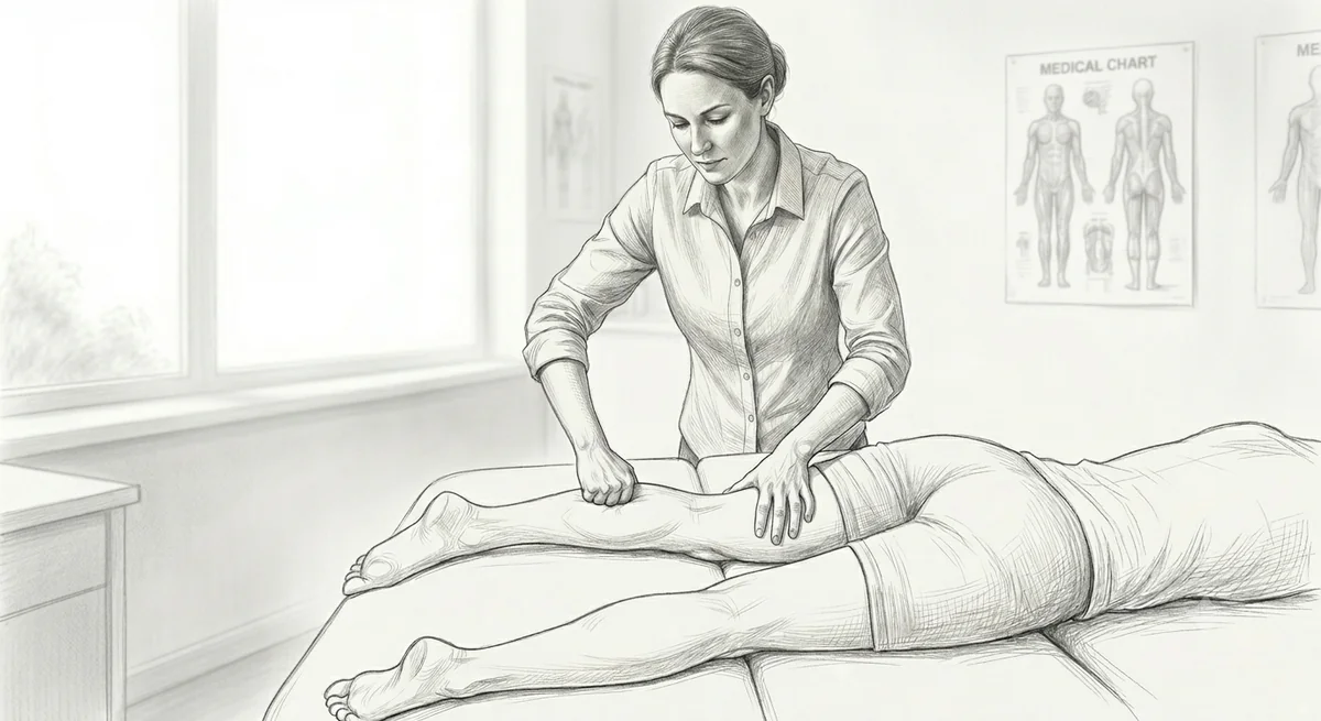
What is fascia?
Fascia is a type of connective tissue that permeates your entire body. It surrounds, supports, protects, connects, and divides your muscles, bones, organs, nerves, and blood vessels — creating a continuous three-dimensional web from head to foot.
A useful analogy: if you imagine an orange, fascia is similar to the white pith — the material under the skin (the rind) and in between each segment of the orange. Like the pith, fascia holds everything together, gives each part its shape, and allows the whole to function as a coordinated unit.
Unlike the orange pith, however, your fascia is dynamic. It slides and glides between layers, responds to movement and load, houses sensory receptors that help coordinate muscle action, and changes its mechanical properties in response to how you use your body. Lubrication is central to all of this — and fascial dysfunction begins when lubrication is reduced.
The three main types of fascia
| Superficial | The layer just below your skin and above your deep fascia, sandwiched between two layers of fat. It cushions and protects nerves and blood vessels close to the surface. |
| Deep | A continuous layer running from the trunk through to the upper and lower limbs. This is the layer most relevant to musculoskeletal pain and the primary focus of Fascial Manipulation — it is above your muscles, below your skin and superficial fascia, and is considered the key structure for transmitting forces across the body. |
| Visceral | The fascial tissue that surrounds and supports your internal organs, providing cushioning and maintaining their position within the body cavities. |
What the deep fascia actually does
The deep fascia is far more than a passive wrapping. It plays three active roles that matter directly to movement and pain:
Force transmission. Your deep fascia is connected to the underlying muscles via direct myofascial attachments. These connections allow it to behave like a long-distance tendon — transmitting muscular forces across multiple joints and body regions simultaneously. The latissimus dorsi on one side, for example, connects through the thoracolumbar fascia to the gluteus maximus on the other. When this system works well, the body moves efficiently. When it is restricted, the load concentrates in structures — joints, tendons, discs — that were not designed to absorb it alone.
Energy storage and release. Deep fascia can absorb, store, and release kinetic energy like a spring. This elastic function makes movement more efficient, with less muscular effort required. Rotational movements — running, walking, throwing — rely heavily on this mechanism. Restricted fascia reduces this efficiency and increases the muscular demand of everyday movement.
Sensory coordination. The deep fascia is home to a dense network of sensory receptors — mechanoreceptors that detect movement, pressure, and tissue tension. This information is processed largely subconsciously and used to coordinate muscle timing, reaction speed, balance, and the appropriate resting tone of muscles throughout the body. When the fascia is densified, this sensory information is degraded, and the coordination it supports begins to break down.
"The thoracolumbar fascia appears to be a structure well-suited to transferring loads between the upper and lower extremities."
Willard et al. (2012). Journal of Anatomy.Hyaluronan — the lubricant your fascia depends on
Between the layers of fascia, and throughout the loose connective tissue that allows them to glide, sits a molecule called hyaluronan (also written as hyaluronic acid). It is a naturally occurring polysaccharide with an extraordinary capacity to bind water — enabling it to act as a highly effective lubricant between moving tissue layers.
Hyaluronan is found throughout the body — in joints, skin, eyes, and connective tissues — but its role in the loose connective tissue between fascial layers is particularly important for musculoskeletal function. Under normal conditions, it exists in a low-viscosity, fluid state that allows the fascial layers to slide easily relative to each other during movement.
This sliding is not incidental. It is the mechanism by which the sensory receptors housed within the fascia are activated. And it is what allows multiple muscle groups to work together as a coordinated system rather than as isolated units.
A specialist cell type called a fasciacyte — identified relatively recently — appears to regulate the production and distribution of hyaluronan within the fascial layers. When these cells are disrupted by sustained compression, repeated microtrauma, or chemical changes in the local tissue environment, the hyaluronan they produce can change its properties in ways that impair fascial gliding.
When dysfunction occurs: densification
When fascial dysfunction develops, hyaluronan undergoes a state change — shifting from its normal fluid, sol-like form toward a denser, more viscous gel. In this altered state, it begins to bind to itself rather than to water, aggregating into clusters that thicken the space between fascial layers and reduce their ability to glide.
This process is called densification. It is important to understand that densification is not the same as scarring or fibrosis — there is no permanent structural change to the tissue. It is a change in the fluid state of the loose connective tissue, and under the right conditions, it is reversible.
Densification reduces the normal gliding between fascial layers. This has two consequences:
Sensory degradation. The mechanoreceptors within the fascia cannot be activated normally when the tissue is stiff and cannot slide. Coordination, proprioception, and movement accuracy all suffer as a result — often subtly, which is why people are frequently unaware that this is part of their problem.
Pain sensitisation. The stiffened tissue increases the sensitivity of the free nerve endings that detect painful stimuli. Movements or positions that would not normally provoke pain begin to do so — and the threshold for pain remains lowered as long as the densification persists.
Densification can develop as a result of trauma, sustained compression (such as prolonged sitting or repetitive posture), overuse or underuse of a region, inflammation, changes in tissue pH, or changes in local temperature. It often accumulates gradually, well before symptoms appear, which is why treatment sites frequently differ from the location of pain.
"Hyaluronan can change its properties in densified fascia, shifting toward a more viscous state that impairs the normal gliding between fascial layers."
Stecco A et al. (2023). Life. | Pratt RL (2021). International Journal of Molecular Sciences.Research also suggests that the fascial environment exists on a spectrum — from reversible densification at the functional end to measurable structural thickening and, in extreme cases, frank fibrosis at the other. A surgical study of patients undergoing occipital nerve decompression for intractable migraine found that 94% had trapezius fascia thicker than 3 mm and fibrotic at surgery (Gfrerer et al., 2020). These are findings at the severe end of the spectrum, not what is typical in general musculoskeletal care — but they illustrate why tissue quality matters. Read more about the densification-to-fibrosis continuum.
How Fascial Manipulation works
The assessment process in Fascial Manipulation by Stecco (FM) uses a systematic analysis of movement and palpation to identify which fascial points — called centres of coordination — are densified and are contributing to the movement pattern that is loading the symptomatic structure. The treatment site is often remote from the site of pain: a densified point in the gluteal fascia, for example, may be contributing to knee pain by altering how load is transmitted through the posterior chain.
Treatment involves sustained, precise manual pressure applied to the identified centre of coordination. The pressure generates both compression and shear forces within the loose connective tissue, and this produces a localised increase in tissue temperature. The combination of shear force and heat creates the conditions for the densified hyaluronan to return from its gel-like state toward its normal, more fluid consistency — restoring the gliding capacity of the fascial layers.
This is the mechanism through which treatment aims to restore normal fascial mechanics. As gliding is restored, sensory receptor function begins to normalise, the abnormal load distribution that was driving the symptomatic structure is reduced, and the pain sensitivity associated with the densified tissue tends to decrease.
The treatment itself — sustained manual pressure to a small area of dense tissue — is characteristically uncomfortable during application. The sensation reflects the nociceptive character of deep fascial tissue: experimental stimulation of the fascia in human volunteers consistently produces pain described as heavy, burning, or agonizing — distinct from the dull, deep ache more typical of muscular stimulation, and often broader in its distribution. This resolves quickly after treatment ends, and any local soreness in the 24–48 hours that follow reflects the tissue-level processes associated with hyaluronan restoration, not tissue damage.
"Fascial Manipulation produced measurable changes in T1ρ MRI signal in treated tissue areas — providing objective evidence of altered tissue composition following treatment."
Menon RG et al. (2020). International Journal of Environmental Research and Public Health.MRI evidence using T1ρ imaging has confirmed that Fascial Manipulation produces measurable changes in fascial tissue composition at treated sites — providing objective support for the proposed mechanism of action. This research, published in the International Journal of Environmental Research and Public Health, is the first to capture structural change in fascia following manual therapy using imaging. Read the full analysis of this research.
Think this might be what's going on for you?
Book an assessmentWho developed this method?
Luigi Stecco (born 1949) is an Italian physiotherapist who spent more than four decades developing what would become Fascial Manipulation by Stecco — beginning in the 1970s with a fundamental dissatisfaction with the techniques available to him. His own description of the problem: existing manual therapy was "not effective enough — some was even useless." That dissatisfaction drove a career-long investigation.
His method did not emerge from a single insight. It was built gradually, drawing from a range of earlier traditions: Elisabeth Dicke's connective tissue massage (1920s Germany), James Cyriax's transverse friction massage, the kinetic chain theories of Françoise Mézières and Herman Kabat, Ida Rolf's structural integration, the trigger point work of Travell and Simons, and early observations about acupuncture meridians. What connected these threads was cadaveric dissection — extensive, systematic, and collaborative — that allowed Luigi to verify anatomical structures and their relationships directly rather than theorise about them.
His early working name for the technique was the neuro-connective technique. He originally believed results came from freeing nerves within connective tissue — an idea that has since been formalised in the literature as fascial entrapment neuropathy. As the anatomical picture became clearer, the model shifted toward the loose connective tissue between fascial layers, the role of hyaluronan, and the concept of centres of coordination — and the name became Fascial Manipulation.
"Manus sapiens, potens est."
A knowledgeable hand is a powerful hand — Luigi Stecco's guiding philosophy and the FM motto.Luigi's daughter, Carla Stecco, is a medical doctor and anatomist at the University of Padua who has led the anatomical science underpinning the method. Her Functional Atlas of the Human Fascial System (2015) was the first systematic anatomical atlas to map fascial connections across the whole body and propose a rational nomenclature for both structural and functional aspects of fascia. The research from Padua — on fasciacytes, hyaluronan, densification, and fascial innervation — forms the evidence base that distinguishes FM from the wider field of soft tissue therapy.
Luigi's early Italian publications reached the English-speaking world in part through practitioners who sought him out before the method was widely known outside Europe. Julie-Ann Day, an Australian physiotherapist who relocated to Italy, was among the first English-speaking practitioners to encounter Luigi's work — meeting him in the early 1990s, before his method had been formally translated. She went on to translate his publications, contribute to the research literature, and has remained one of the longest-serving members of the FM teaching faculty. She is a co-author, alongside Carla and Antonio Stecco, of a 2009 clinical study on FM applied to chronic shoulder pain — one of the early published studies on the method in English.
Dr Steven Hewitt trained in Fascial Manipulation under Julie-Ann Day. That lineage runs from Luigi's original clinical work, through the Padua research group, directly to the approach used at Elevate Health Care.
FM is now taught internationally through the Fascial Manipulation Association. Practitioner certification involves multiple levels of practical and written examination.
How FM differs from massage, dry needling, and physiotherapy
Several manual therapy approaches address soft tissue, and people often ask how Fascial Manipulation relates to them. The differences are meaningful — not because other approaches are ineffective at what they are designed to do, but because they are designed to do different things.
The table below describes what each approach primarily targets and how assessment and treatment are structured. It is not a comparison of outcomes — individual presentations, practitioners, and clinical contexts vary considerably, and many people receive more than one type of treatment over the course of their care.
Swipe to compare all four approaches →
| Remedial / sports massage | Dry needling | General physiotherapy | Fascial Manipulation (Stecco) | |
|---|---|---|---|---|
| Primary target | Superficial and deep muscle tissue — tension, circulation, recovery | Trigger points within muscle fibres — localised taut bands producing referred pain | Varies by presentation and practitioner — may include joint, muscle, nerve, or movement patterns | The loose connective tissue between fascial layers — where densification of hyaluronan has reduced gliding and altered sensory coordination |
| Assessment | Clinical interview; symptom-based palpation of tense or tender tissue | Palpation of taut bands; identification of referred pain patterns | Varies — typically combines history, movement testing, orthopaedic and/or neurological testing | Systematic 4-step protocol: case history, movement verification across 6 directions per segment, palpatory verification of centres of coordination, then treatment |
| Treatment site | At or near the site of pain or perceived tension | At the trigger point — typically at or near the pain site | At or near the symptomatic structure | At centres of coordination — specific fascial points identified through palpation, often remote from the pain site |
| What determines the site | Symptom location; palpation of tender tissue | Trigger point maps; referred pain patterns | Clinical presentation and diagnostic testing | Palpatory density of fascial tissue — the degree of densification, not the pain report alone |
FM is not the only approach that looks beyond the site of pain — some physiotherapy and dry needling models do this too. What is specific to FM is the anatomical model it works from (the fascial system as a force-transmission and sensory network), the assessment method (which prioritises mechanical palpation of fascial tissue over pain location), and the treatment target (the loose connective tissue between fascial layers rather than the muscle, joint, or nerve directly).
These approaches are not mutually exclusive. FM is sometimes one component of a broader management plan that includes exercise, movement retraining, or other therapeutic input.
For a fuller side-by-side look at how Fascial Manipulation relates to remedial massage, dry needling, physiotherapy and chiropractic adjustments, see How Does Fascial Manipulation Compare?
What to expect
-
BeforeAssessment determines everything No treatment is applied without first completing the full FM assessment — Interview, Movement Verification, and Palpatory Verification. This four-step process identifies which specific points in the fascial system are densified, which movement directions are restricted, and how these relate to your pain. The assessment takes time and is not abbreviated. Treatment follows directly from what it reveals — not from where you are reporting pain.
-
DuringPrecise, sustained pressure — often not where you expect Treatment is applied to specific centres of coordination in the deep fascia — small, precise areas identified through palpatory verification. These points are frequently remote from your area of pain. This is expected and intentional: the densified tissue driving the problem is often not at the painful site. The reason is that altered load sharing is itself clinically silent: when one part of a chain stops taking its share, you do not feel the redistribution — you feel the tissue that ends up carrying the excess. The painful structure is typically the victim, not the culprit. The sensation at the contact point is typically intense — often beginning as a sharp sensation that softens toward a dull ache as the application continues. This reflects how sensitised the densified tissue has become: pressure that would feel mild over healthy fascia produces a significantly amplified response at a densified point. The intensity at each point generally reduces as treatment progresses, and is usually considerably diminished by the time the practitioner moves on. It is distinctly uncomfortable but tolerable, and the arc from sharp to dull is itself a clinical indicator that the correct tissue is being engaged. A typical session addresses three to six points depending on the complexity of the presentation, with sustained pressure applied to each for two to four minutes.
-
AfterAvoid anti-inflammatories for 48–72 hours This is the most practically important post-treatment instruction. We generally recommend avoiding anti-inflammatory medication — ibuprofen (Nurofen), naproxen, diclofenac (Voltaren) and similar — where possible, for 48–72 hours after treatment. The reasoning is that treatment initiates a hyaluronan restoration process involving a brief local inflammatory response at the treated sites, and the current understanding is that anti-inflammatory medication may dampen that response. Some local soreness at treated points is normal and expected for 24–48 hours — this reflects the HA gel-to-sol transition and associated tissue activity, not damage. Staying well hydrated and keeping moving lightly in the days following treatment supports recovery, and if the post-treatment ache is too uncomfortable, paracetamol is an alternative analgesic that is not thought to affect the treatment response in the same way. None of this applies to medication you have been prescribed. Never stop or change prescribed medication — including low-dose aspirin taken for heart or stroke prevention — without speaking to your GP or pharmacist.
-
ResponseResponses vary — and may not be where you expect them Some people notice an immediate change in mobility or pain following the first session. Others notice a gradual shift over 24–72 hours as the tissue changes settle. It is not unusual to notice improvement in an area different from where treatment was applied — this reflects the fascial chain connections, not an unexpected outcome. Complex or long-standing presentations typically require a series of sessions before the full picture of change is established; three to six sessions is a common clinical course, with reassessment built in throughout. Steve will discuss realistic expectations with you at your initial assessment based on the specific pattern of densification identified.
whether this applies to you?
We have availability.
References
- PubMed Willard FH, Vleeming A, Schuenke MD, Danneels L, Schleip R. (2012). The thoracolumbar fascia: anatomy, function and clinical considerations. Journal of Anatomy, 221(6), 507–536.
- PubMed Stecco C, Stern R, Porzionato A, et al. (2011). Hyaluronan within fascia in the etiology of myofascial pain. Surgical and Radiologic Anatomy, 33(10), 891–896.
- PubMed Pratt RL (2021). Hyaluronan and the fascial frontier. International Journal of Molecular Sciences, 22(13), 6845.
- PubMed Stecco A, Bonaldi L, Fontanella CG, Stecco C, Pirri C. (2023). The effect of mechanical stress on hyaluronan fragments' inflammatory cascade: clinical implications. Life, 13(12), 2277.
- PubMed Menon RG, Oswald SF, Raghavan P, Regatte RR, Stecco A. (2020). T1ρ-mapping for musculoskeletal pain diagnosis: case series of variation of water bound glycosaminoglycans quantification before and after fascial manipulation in subjects with elbow pain. International Journal of Environmental Research and Public Health, 17(3), 708.
- PubMed Weiss K, Kalichman L. (2021). Deep fascia as a potential source of pain: a narrative review. Journal of Bodywork and Movement Therapies, 28, 82–86.