Ready to get on top of this?
📞 Call Now — speak with our team
🗓 Book Online — available 24/7
What Is the Deep Longitudinal Sling?
The deep longitudinal sling is a posterior chain of anatomical structures that connects the posterior lower leg to the lumbar spine. Running continuously along the back of the body, it includes:
- Peroneal muscles (fibularis longus and brevis) — at the lateral lower leg and ankle
- Biceps femoris (long head) — the lateral hamstring, running from the ischial tuberosity to the fibular head
- Sacrotuberous ligament — one of the primary passive restraints of the sacroiliac joint, connecting the ischial tuberosity to the lateral sacrum and coccyx
- Deep lamina of the thoracolumbar fascia (TLF) — arising from the erector spinae and connecting into the sacrotuberous ligament above
- Erector spinae and multifidus — the deep lumbar extensors, whose aponeuroses form the deep lamina of the TLF
The sling is not a single structure — it is a functionally continuous chain. The muscle and connective tissue elements are not mechanically isolated from each other: force generated at one end of the chain is transmitted across the whole length. Van Wingerden, Vleeming and colleagues (1993) were among the first to demonstrate directly that tension applied to the biceps femoris places measurable load on the sacrotuberous ligament — establishing the mechanical basis for the lower limb–to–pelvic ring connection that defines the deep longitudinal sling. Subsequent in-vivo research has confirmed that restriction in the ankle and lower leg is capable of altering soft tissue tension through the dorsal thigh — further illustrating how mechanically integrated this chain is in the living body (Wilke et al., 2020).
Orientation and Plane of Action
The deep longitudinal sling is primarily a sagittal plane structure. Its primary function is to resist and manage forces acting in the forward-backward direction — particularly the anterior shear forces that occur at the lower lumbar vertebrae and sacrum during walking, running, and lifting.
| Component | Level | Connection |
|---|---|---|
| Erector spinae / multifidus | Lumbar | Forms TLF deep lamina |
| TLF deep lamina | L4–S1 | Connects to sacrotuberous ligament |
| Sacrotuberous ligament | Sacrum/ischial tuberosity | Key SIJ passive restraint |
| Biceps femoris (long head) | Ischial tuberosity → fibular head | Primary hamstring contributor |
| Peroneal chain | Fibular head → lateral ankle | Continues the chain to the foot |
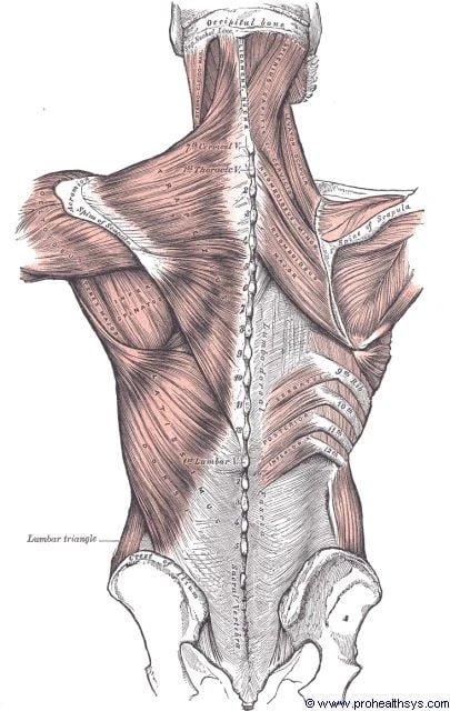
The thoracolumbar fascia (broad white band, lumbar region) — the connective tissue hub through which the deep longitudinal sling operates. The erector spinae and multifidus form the deep lamina of the TLF, which connects downward to the sacrotuberous ligament and into the biceps femoris. Load transferred up from the posterior lower limb via this chain enters the TLF here before being distributed across the lumbar spine. Illustration: Henry Vandyke Carter, Gray's Anatomy (1918) — public domain.
What Does It Do?
Anti-Shear Load Transfer During Gait
During walking and running, each stride creates a forward shear force on the lower lumbar vertebrae — the upper body moves forward while the lower limb drives back and pushes off. The deep longitudinal sling is one of the primary mechanisms by which this shear force is shared across the posterior chain, rather than being left to the disc and facet joints alone.
As the leg extends behind the body in the late stance phase of gait, the biceps femoris is loaded under tension. Van Wingerden et al. (1993) demonstrated that this biceps femoris tension is directly transmitted through the sacrotuberous ligament, stiffening the posterior pelvic ring and compressing the sacroiliac joint. That muscular and ligamentous compression is what is meant by force closure — the active contribution to joint stability, as distinct from form closure, which is the passive stability the joint surfaces provide by their shape alone. The sling loads the deep lamina of the TLF at the same time, bracing the lower lumbar segments.
A well-functioning deep longitudinal sling means each step is cushioned and load-shared across a long chain. A restricted or dysfunctional sling means each step concentrates more stress at the lumbar spine and SIJ.
Sacrotuberous Ligament Pre-Load and SIJ Stability
The sacrotuberous ligament is one of the passive stabilisers of the sacroiliac joint — it resists sacral nutation (the forward rotation of the top of the sacrum) and helps to close the joint against the compressive forces of the body weight above. The deep longitudinal sling applies tensile load to this ligament during movement, contributing to its passive stiffening effect on the SIJ. Aldabe et al. (2019), in a systematic review of the sacrotuberous ligament's anatomy, biomechanics, and clinical relevance, confirmed the STL's role as a primary posterior SIJ passive restraint and described its mechanical integration with the surrounding musculofascial structures — including its direct connections to the biceps femoris, the gluteus maximus, and the TLF. Kim et al. (2024) further characterised the histomorphological continuity between the sacrotuberous ligament and the hamstring origins, providing tissue-level evidence for the myofascial integration of the lower limb and the posterior pelvic ring at this junction.
When the deep longitudinal sling is restricted — through hamstring tightness, TLF densification, or reduced ankle mobility — this pre-load effect is reduced and the SIJ is relatively unsupported.
Connection to the Foot
The inclusion of the peroneal muscles in the chain means the deep longitudinal sling extends all the way to the lateral ankle and foot. This is clinically significant: a history of ankle sprain, chronic ankle instability, or restricted ankle dorsiflexion can alter the mechanics of the entire chain upward — contributing to hamstring loading, reduced sacrotuberous ligament tension, and increased lumbar spine shear. This is one reason why old ankle injuries are sometimes relevant to recurrent lower back problems.
The chain does not stop at the ankle either. Dissection and MRI work has shown that the plantar fascia is continuous with the paratenon of the Achilles tendon around the calcaneus, rather than the two simply meeting at the heel bone (Stecco et al., 2013). That continuity places the sole of the foot at the bottom of the same posterior line, and it is why heel and Achilles presentations are not necessarily local problems: tension arriving from above has somewhere to go, and restriction below has somewhere to travel. It is also why we look at the hip and lumbopelvic end when someone presents with plantar fasciopathy or Achilles tendinopathy that has not settled with local loading alone.
When It Goes Wrong: Clinical Relevance
Tight Hamstrings and the Posterior Chain
The most common point of restriction in the deep longitudinal sling is the biceps femoris and the posterior thigh. Chronic hamstring tightness — whether from prolonged sitting, athletic overuse, or post-injury adaptive shortening — places the sling in a state of chronic tension that reduces the compliant force-transmitting function of the chain and concentrates compressive load at the sacrotuberous ligament and sacroiliac joint.
Importantly, isolated hamstring stretching often provides only temporary relief in this pattern — because the restriction is not only in the muscle belly itself, but in the fascial environment surrounding the chain. When the deep fascia overlying the hamstrings and the fascial connections into the sacrotuberous ligament are restricted, stretching the muscle generates tension through the system without restoring the gliding between layers that allows the chain to function smoothly.
The Desk Worker Pattern
Prolonged sitting places the hamstrings in a shortened position, the pelvis in anterior tilt, and the lower lumbar spine in sustained extension. Over time, this posture progressively stiffens the deep longitudinal sling in a shortened state — reducing the available posterior chain length for functional movement and loading the lower lumbar facet joints and SIJ with the residual compressive effect.
We commonly see this pattern in desk workers who also exercise regularly: someone who sits for eight hours and then runs or lifts weights in the evening. The deep longitudinal sling is already operating in a restricted, shortened state, and the exercise load is applied without first restoring the chain's compliance. This is a common contributor to recurrent lower back and SIJ pain in active desk workers.
The Runner and the Lateral Chain
In runners, the deep longitudinal sling is loaded heavily at the push-off and late stance phases of gait. Restriction in the biceps femoris, or a history of ankle injury that has altered foot mechanics, commonly alters the loading through the lateral chain — producing asymmetric tension at the sacrotuberous ligament, which over time contributes to unilateral SIJ irritation, L5/S1 facet loading, or lateral hamstring injury.
Previous Ankle Injuries
The peroneal component of the deep longitudinal sling means that old lateral ankle sprains are genuinely relevant to posterior chain function. Ankle sprains cause fascial restriction and altered mechanoreceptor function around the ankle and peroneal compartment. Research has demonstrated that ankle motion is associated with soft tissue displacement in the dorsal thigh — confirming that the ankle and posterior thigh are mechanically linked in the living body (Wilke et al., 2020). A clinical picture of recurrent lower back pain in someone with a history of multiple ankle sprains is one in which assessment of the deep longitudinal sling is a logical and specific step.
The Fascial Lens: Why We See This Differently
The deep longitudinal sling is not simply a chain of muscles. The continuity of the chain depends entirely on the fascial structures that connect the muscular elements — the TLF, the sacrotuberous ligament, the bicipital fascia and hamstring fascia, the crural fascia of the lower leg.
When the fascial layers within any component of this chain become densified — that is, when the hyaluronan-rich loose connective tissue between the fascial layers increases in viscosity and loses its normal gliding capacity — the functional continuity of the sling is disrupted. The muscles can still contract, but the force they generate is not transmitted smoothly across the chain. Abnormal load sharing during movement may contribute to overload of one tissue in particular, and it is the overloaded tissue the patient feels. The altered load sharing itself is clinically silent.
This is clinically important for two reasons:
1. The restriction may not be where the pain is. TLF densification at the L5-S1 level can restrict the sacrotuberous ligament's ability to be loaded from the biceps femoris above — but the patient's pain is in the lower back, not in the hamstring or TLF. Conversely, restriction in the peroneal fascia from an old ankle injury can alter the mechanics of the entire posterior chain upward — but the patient presents with lumbar or SIJ pain.
2. The restriction may be in the fascia rather than the muscle. A patient who has diligently stretched their hamstrings for years without resolution of posterior chain tightness is a common presentation. The myofascial tension that persists despite regular stretching is frequently a fascial densification issue — the layers surrounding the chain are restricted, and stretching the muscle alone does not address the inter-layer gliding problem.
Luomala and Pihlman (2017) explain this through the Fascial Manipulation model: the retromotion (RE) sequence — which corresponds anatomically to the deep longitudinal sling — is assessed through the Centres of Coordination (CCs) of each body segment along the posterior chain. Palpatory verification identifies which specific points within the chain are most densified and tender. Treatment restores gliding at those points rather than applying generalised posterior chain stretching.
The Fascial Picture — Deep Longitudinal Sling
The deep longitudinal sling functions as a continuous load-transmitting chain. Its effectiveness depends not only on the strength and length of its muscular components, but on the gliding capacity of the fascial layers that connect them. Densification anywhere in the chain — at the TLF, the sacrotuberous ligament, the hamstring fascia, or the peroneal compartment — disrupts the whole chain's ability to distribute force. This is why treatment directed at the chain as a whole, including its fascial components, may support more complete resolution than isolated stretching or strengthening.
What Does the Research Say?
The sling is not a drawing. Each link in it has been examined directly, and the evidence divides into three questions.
Is the chain anatomically real? Largely yes, for this sling in particular. A systematic review of myofascial chains found the evidence across the body to be of mixed quality — but the posterior chain corresponding to the deep longitudinal sling drew some of the strongest anatomical support in it. Dissection work has mapped the continuity of the fascial system from the lumbar region through the sacrum and into the posterior thigh, and tissue-level analysis of the junction between the sacrotuberous ligament and the hamstring origin found structural features consistent with genuine continuity rather than two structures merely lying against one another.
Does tension actually travel along it? This is the older and firmer finding. In 1993 van Wingerden and colleagues showed that tension in the biceps femoris measurably stiffens the sacrotuberous ligament — the foundational evidence that the lower limb contributes to posterior pelvic ring stability. More recently the same has been shown in living people rather than cadavers: ankle motion produces measurable soft-tissue displacement in the back of the thigh, which is direct evidence that the lower leg and posterior thigh are mechanically linked.
And does it matter to the lumbar spine? The thoracolumbar fascia is now described as a central load-transferring structure of the lumbar spine whose contribution to stability has been underappreciated, and the sacrotuberous ligament as a multi-attachment structure with direct mechanical links to the biceps femoris, gluteus maximus, piriformis and TLF — loaded both passively by body weight and dynamically by the muscles of this sling. The practical consequence is that a restriction in any of the structures attaching to it can alter how the posterior pelvic ring is loaded.
The eight studies behind this, one by one
Myofascial Force Transmission Across the Chain
Wilke et al. (2018), in a review examining fascia's force transmission capacity, demonstrated that myofascial connections enable force transmission not only within a muscle but across muscles and joints — with fascial chains acting as conduits for load distribution across the body. This work supports the clinical view that restriction at one level of the deep longitudinal sling is capable of altering mechanics at remote sites.
In-Vivo Evidence for the Posterior Chain
Wilke et al. (2020) demonstrated in-vivo that ankle motion was directly associated with soft tissue displacement in the dorsal thigh — providing direct evidence that the lower leg and posterior thigh are mechanically linked through continuous soft tissue connections. This study supports the clinical relevance of ankle function to posterior chain loading and, by extension, to the sacrotuberous ligament and sacroiliac joint mechanics.
The Systematic Review of Myofascial Chains
Wilke et al. (2016) conducted a systematic review examining the anatomical and in-vivo evidence for myofascial chains, concluding that while the evidence is of varying quality, anatomical continuity between posterior chain structures is well-supported. The posterior chain corresponding to the deep longitudinal sling received some of the strongest anatomical support in this review.
The TLF in Spine Biomechanics
Driscoll et al. (2018), reviewing the fascia's role in spine biomechanics, described the TLF as a central load-transferring structure in the lumbar spine — noting that its passive and active contributions to spinal stability have been significantly underappreciated. The deep lamina of the TLF, which is the component most directly implicated in the deep longitudinal sling, contributes substantially to resistance against anterior shear on the lower lumbar vertebrae.
Fascial Anatomy of the Back
Benjamin (2009), in a comprehensive review of the fascia of the limbs and back, mapped the anatomical connections of the TLF and posterior fascial layers in detail — confirming the continuity of the fascial system across the lumbar, sacral and posterior thigh regions, and its clinical relevance to load transfer and pain.
The Original BF–Sacrotuberous Ligament Evidence
Van Wingerden, Vleeming et al. (1993) directly examined the mechanical interaction between the biceps femoris and the sacrotuberous ligament, demonstrating that tension in the biceps femoris produced measurable stiffening of the STL — establishing the foundational biomechanical evidence for the lower limb contribution to posterior pelvic ring stability that underpins the deep longitudinal sling concept. This work preceded the wider adoption of the myofascial sling framework and remains one of the most directly relevant studies for understanding how hamstring function and SIJ stability are mechanically coupled.
The Sacrotuberous Ligament: Systematic Review
Aldabe et al. (2019) conducted a systematic review of the sacrotuberous ligament's anatomy, mechanical properties, and clinical relevance, identifying it as a multi-attachment structure with direct mechanical links to the biceps femoris, the gluteus maximus, the piriformis, and the TLF. The review confirmed that the STL is loaded both passively (through body weight and sacral nutation) and dynamically (through the muscular components of the deep longitudinal sling during movement) — and that restriction in any of the structures connecting to the STL has the potential to alter posterior pelvic ring mechanics.
Histomorphological Evidence at the STL–Hamstring Junction
Kim et al. (2024) examined the histological and morphological characteristics of the junction between the sacrotuberous ligament and the hamstring origin, finding structural features consistent with myofascial continuity across this junction. The findings provide tissue-level evidence for the mechanical integration of the hamstring chain and the posterior pelvic ring — supporting the clinical relevance of hamstring restriction and fascial densification in the posterior thigh to sacrotuberous ligament loading and SIJ function.
How We Assess and Address This
Our assessment of the deep longitudinal sling is designed to evaluate the chain across its full length — not only at the painful site:
- Posterior chain length assessment — active and passive straight-leg raise, hamstring extensibility, and modified Thomas test to assess the contribution of hip flexor restriction to posterior chain loading
- Ankle dorsiflexion assessment — restricted ankle DF is a consistent finding in restricted deep longitudinal sling function; we assess both the talocrural mobility and the fascial compliance of the peroneal compartment
- Sacrotuberous ligament palpation and loading assessment — direct assessment of the posterior pelvic ring, including sacrotuberous ligament tension and SIJ mobility
- TLF fascial assessment using Stecco FM protocols — palpatory verification of the retromotion (RE) sequence CCs from the lumbar to the ankle; identifying which levels of the chain have the greatest fascial restriction
- Lumbar and SIJ movement assessment — to identify the functional consequences of deep longitudinal sling restriction at the spinal and pelvic level
Treatment is directed at the fascial environment of the chain and the underlying movement patterns:
- Fascial Manipulation by Stecco — targeted manual therapy at the most densified CCs along the retromotion (RE) sequence, from the lumbar TLF through the posterior thigh to the peroneal compartment
- Graduated posterior chain rehabilitation — restoring posterior chain length and progressive loading through the chain under controlled conditions, including Nordic hamstring work, Romanian deadlift variations, and single-leg posterior chain patterns
- Ankle mobility work — where ankle dorsiflexion is restricted, specific ankle mobility work is incorporated to restore the full chain's functional length
- Addressing the desk-worker pattern — hip flexor mobility, postural load management, and movement pattern correction where anterior pelvic tilt is contributing to posterior chain shortening
New to Fascial Manipulation? Read how it works →
Please note: The information on this page describes our general clinical approach and is intended for educational purposes only. Individual presentations vary, and your assessment and management will be tailored specifically to you. Nothing on this page constitutes clinical advice for your individual situation. Please consult a registered health practitioner for advice about your specific condition.
Related Conditions
The deep longitudinal sling is relevant across a wide range of low back and lower limb conditions. If you have been diagnosed with or are experiencing any of the following, deep longitudinal sling assessment may be a useful part of your evaluation:
→ Sacroiliac Joint Syndrome — the sacrotuberous ligament is a central SIJ stabiliser loaded by this sling
→ Lumbar Facet Syndrome — deep longitudinal sling restriction increases posterior facet loading
→ Lumbar Disc Problems — reduced anti-shear capacity of the sling increases disc loading at lower lumbar levels
→ Thoracic Facet & Costovertebral Dysfunction — the paraspinal compartment and the deep lamina of the TLF run continuously from the lumbar region into the thoracic spine, so restriction above changes how this chain loads below
→ Understanding the Posterior Oblique Sling — the posterior oblique sling works in functional partnership with the deep longitudinal sling at the sacrum and TLF
Take the Next Step
The deep longitudinal sling is one of the structures we routinely assess in any presentation of lower back, sacroiliac, or recurrent posterior thigh pain — particularly when isolated treatment of the painful structure has not produced durable improvement. If your pain is in the lower back or pelvis but you have a history of hamstring issues or ankle injuries, a specific assessment of this chain may reveal a contributing pattern that has not previously been identified.
The same applies from the bottom of the chain up. This sling runs from the sole of the foot to the lumbar spine, so a heel, Achilles, calf or hamstring problem that keeps returning is worth assessing along its whole length rather than only where it hurts. Recurrent plantar fasciopathy, a stubborn Achilles tendinopathy, an ankle that never fully settled after a sprain, or proximal hamstring pain that returns whenever load goes back up can each reflect a chain that is not sharing load evenly — and in each case the restriction limiting it may sit well away from the symptoms.
Ready to get on top of this?
📞 Call Now — speak with our team
🗓 Book Online — available 24/7
Located at Suite 30, Level 1, 93 Wells Road, Chelsea Heights, Melbourne. No referral is required — allow 60 minutes for your first appointment.
References
- PubMed Van Wingerden JP, Vleeming A, Snijders CJ, Stoeckart R (1993). A functional-anatomical approach to the spine-pelvis mechanism: interaction between the biceps femoris muscle and the sacrotuberous ligament. European Spine Journal, 2(3), 140–144.
- PubMed Aldabe D, Hammer N, Woodley SJ et al. (2019). A systematic review of the morphology and function of the sacrotuberous ligament. Clinical Anatomy, 32(3), 396–407.
- PubMed Kim M, Yang HM, Yeo IS (2024). Anatomical study of the sacrotuberous ligament and the hamstring muscles: a histomorphological analysis. Clinical Anatomy, 37(4), 383–389.
- PubMed Wilke J et al. (2018). Not merely a protective packing organ: a review of fascia and its force transmission capacity. Journal of Applied Physiology, 124(1), 234–244.
- PubMed Wilke J et al. (2020). Ankle motion is associated with soft tissue displacement in the dorsal thigh: an in vivo investigation suggesting myofascial force transmission across the knee joint. Frontiers in Physiology, 11, 180.
- PubMed Wilke J et al. (2016). What is evidence-based about myofascial chains? A systematic review. Archives of Physical Medicine and Rehabilitation, 97(3), 454–461.
- PubMed Driscoll M (2018). Fascia — the unsung hero of spine biomechanics. Journal of Bodywork and Movement Therapies, 22(1), 90–91.
- PubMed Benjamin M (2009). The fascia of the limbs and back — a review. Journal of Anatomy, 214(1), 1–18.
- Luomala T, Pihlman M (2017). A Practical Guide to Fascial Manipulation. Elsevier.
- PubMed Stecco C, Corradin M, Macchi V et al. (2013). Plantar fascia anatomy and its relationship with Achilles tendon and paratenon. Journal of Anatomy, 223(6), 665–676.