Chiropractic adjustments, the short version
A chiropractic adjustment (more precisely, spinal manipulative therapy) is a quick, controlled, low-amplitude movement applied to a specific joint that's become restricted. It takes a fraction of a second. The pop you sometimes hear is harmless — it's a gas bubble forming in the joint fluid, not bones cracking — and the technique works just as well without it.
The effects are largely neurological, not mechanical — it isn't about putting a bone "back in place." At this clinic, adjustment is one tool, used only when the assessment calls for it — often alongside or after fascial treatment.
A chiropractic adjustment is a specific, targeted manual technique — not a generic intervention applied to the whole spine. At Elevate Health Care, adjustments are used when movement analysis and palpatory assessment identify a joint that is contributing to the mechanical picture. They are one component of a broader clinical approach that also addresses the fascial system and movement patterns — not a standalone treatment, and not applied to every patient or every visit.
What is a chiropractic adjustment?
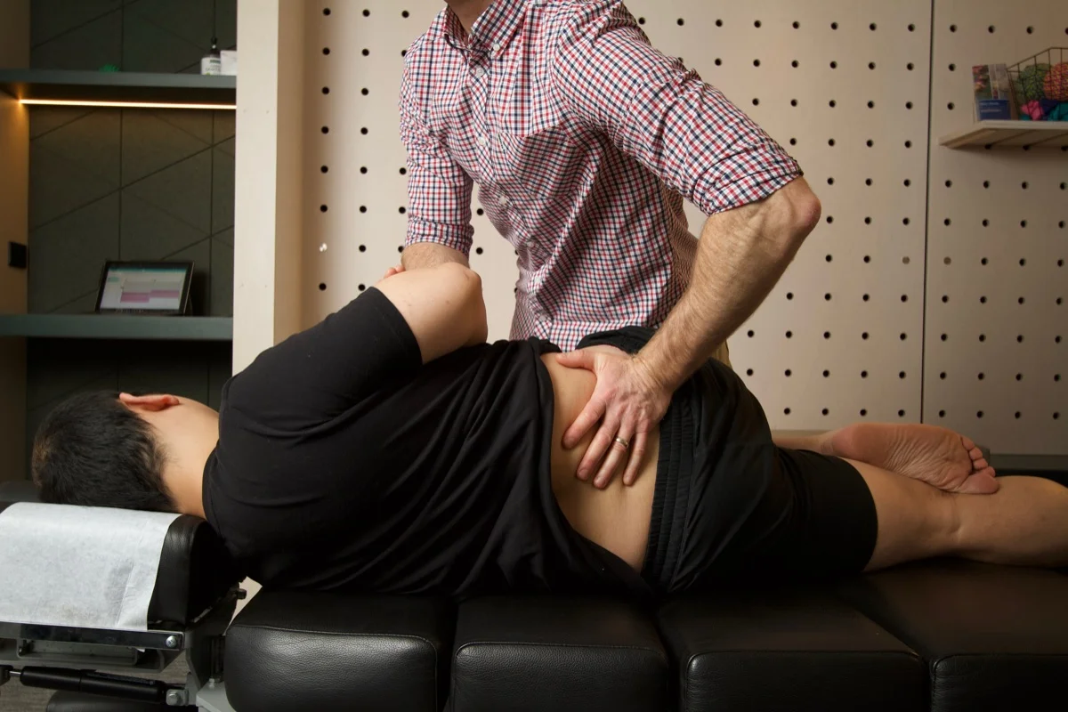
A chiropractic adjustment — more precisely, a spinal manipulative therapy (SMT) or high-velocity, low-amplitude (HVLA) thrust — is a controlled manual technique applied to a specific spinal or extremity joint. The practitioner positions the joint at or near the end of its available range, then applies a precise, brief force through a specific vector.
The total duration of the thrust is typically measured in milliseconds — far too fast to be a stretch, and too brief for the surrounding muscles to generate a significant protective response. The technique requires a precise contact point, a specific patient position, a particular direction of force, and controlled depth. These variables are not interchangeable — they are what makes a skilled adjustment different from a generic mobilisation.
The goal is to restore normal joint motion at a segment that has become restricted — and, through the neurological effects of the technique, to reduce the aberrant sensory input from that joint that is influencing muscle tone and pain sensitivity in the surrounding region.
Adjustments are applied to the spinal joints (cervical, thoracic, and lumbar) and to peripheral joints including the hip, shoulder, elbow, wrist, ankle, and foot — depending on what the assessment reveals.
What is the sound?
The audible pop or crack that sometimes accompanies a chiropractic adjustment is one of the most commonly misunderstood aspects of the technique. It is not bones rubbing together, not the spine cracking, and not an indication that anything has been broken or damaged.
The sound is a cavitation event within the synovial fluid of the joint. Synovial joints contain a small amount of synovial fluid, which in turn contains dissolved gases — primarily carbon dioxide and nitrogen. When the joint surfaces are rapidly distracted by the thrust, the pressure within the joint cavity drops momentarily. This causes dissolved gas to come out of solution rapidly, forming a gas bubble. The audible pop is the sound of that bubble forming — not bursting.
Following cavitation, there is a brief refractory period during which the same joint cannot be made to cavitate again. The gas gradually re-dissolves into the synovial fluid over the following several minutes.
Importantly, the therapeutic effect of an adjustment does not depend on whether cavitation occurs. A technique that achieves the same joint positioning and neurological response without an audible pop is equally valid clinically. The sound is a physical byproduct of the technique — not its mechanism of action.
The proposed mechanisms
Research into why spinal manipulation produces its clinical effects has identified several plausible neurophysiological mechanisms. These are not mutually exclusive — they likely operate simultaneously and to varying degrees depending on the clinical presentation and the technique applied.
-
1Joint mechanoreceptor activation The joint capsule and surrounding tissues contain a dense network of mechanoreceptors — sensory receptors that respond to mechanical deformation, pressure, and movement. The brief, high-velocity thrust of an adjustment activates muscle spindle afferents (Group I and II), Golgi tendon organ afferents, and smaller-diameter Group III/IV fibres in a way that normal voluntary movement cannot replicate. This barrage of proprioceptive input reaches the spinal cord and brain within milliseconds, where it modulates pain signals and alters muscle tone across the surrounding region. [7, 8]
-
2Reflex inhibition of muscle guarding When a joint is restricted or painful, the surrounding muscles commonly enter a state of heightened tone — a protective reflex that itself perpetuates restriction and discomfort. The mechanoreceptor barrage generated by a spinal adjustment can inhibit this guarding response through spinal cord processing, producing a reduction in local muscle hypertonicity that often outlasts the technique. Animal studies demonstrate that spinal manipulation produces both excitatory and inhibitory paraspinal EMG responses — and that in symptomatic patients, the inhibitory response tends to predominate. [8]
-
3Descending pain modulation Spinal manipulation activates descending pain inhibitory pathways — the brain's own system for reducing pain signal intensity. This is mediated through supraspinal structures including the periaqueductal grey (PAG), the rostral ventromedial medulla (RVM), and the anterior cingulate cortex, with downstream release of endogenous opioids and serotonin. The clinical consequence is a measurable reduction in both local and regional pain sensitivity following manipulation — a hypoalgesic effect documented in systematic reviews of experimental pain studies. [7, 9]
-
4Restoration of segmental proprioception Joints restricted in their normal range of movement produce altered sensory input to the central nervous system — information that influences motor control, muscle coordination, and load distribution at adjacent levels. Restoring normal segmental mobility normalises this sensory stream. Research using transcranial magnetic stimulation (TMS) to measure motor cortex excitability has shown that spinal manipulation can alter corticospinal drive — with manipulation producing a 20% reduction in stretch reflex amplitude in patients who experienced joint cavitation, suggesting a resetting of hyperactive spinal reflex pathways. [10]
-
5Reversal of arthrogenic muscle inhibition When a joint is injured, inflamed, or mechanically restricted, aberrant afferent input from that joint actively inhibits the surrounding musculature — a phenomenon known as arthrogenic muscle inhibition (AMI). This is not the same as disuse atrophy. AMI is a neural inhibition of voluntary muscle activation driven by nociceptive and mechanoreceptive signals from the joint itself. It has been documented in the knee, hip, shoulder, and ankle, and explains why muscles that appear intact on imaging can feel weak and unresponsive following joint dysfunction. Restoring normal joint motion — by removing the source of aberrant afferent input — is one mechanism by which manipulation can reduce AMI and restore normal muscle activation patterns. Randomised controlled trial evidence shows that conservative spinal manipulation reduces knee-extensor inhibition in patients with low back and sacroiliac dysfunction. [11, 12]
The relative contribution of each mechanism varies by presentation, technique, and the individual patient's neurological state. Current research supports the view that the effects of spinal manipulation are primarily neurophysiological rather than purely mechanical — the benefit is not simply about moving a bone "back into place." A 2025 overview of 136 studies on the neuromuscular response to high-velocity, low-amplitude manipulation concluded that evidence for EMG, reflex, and proprioceptive changes is growing, though effect sizes and response patterns differ by spinal region and patient population. [13]
What the research says
Spinal manipulation has one of the better evidence bases among manual therapy techniques, particularly for specific presentations. The evidence is strongest for neck pain, cervicogenic headache, and low back pain — and for the use of manipulation within a multimodal approach rather than as a standalone intervention.
"Spinal manipulation is recommended for adults with cervicogenic headache. For tension-type headache, spinal manipulation is recommended within multimodal care."
Trager et al. (2026). Journal of Integrative and Complementary Medicine. Chiropractic Clinical Practice Guideline — CGH & TTH.A 2012 systematic review of manual therapies for cervicogenic headache assessed eight RCTs and found that spinal manipulation and mobilisation produced significant reductions in headache frequency, intensity, and associated disability — with effect sizes comparable to those seen with prophylactic medication. Multimodal approaches combining joint and soft tissue therapy produced superior outcomes to unimodal treatment. [1]
For cervical radiculopathy — arm pain arising from nerve root irritation in the neck — a well-designed RCT found that a combination of manual therapy (including cervical and thoracic manipulation), supervised exercise, and cervical traction produced significantly greater improvements in pain, disability, and patient-perceived recovery at six months compared to home exercise alone: 62% success in the manual therapy group versus 32% in the exercise-only group. [3]
"Spinal manipulation reduced migraine days versus control (Hedges' g = −0.35; p<0.001)."
Rist et al. (2019). Headache. Harvard Medical School & Palmer College of Chiropractic.A Harvard-led systematic review and meta-analysis of six RCTs (pooled n=677) found that spinal manipulation produced a statistically significant reduction in migraine days compared to control conditions — a finding that holds even accounting for the methodological limitations across the included trials. [4]
The 2017 JOSPT Neck Pain Clinical Practice Guidelines recommend thoracic manipulation combined with cervical range-of-motion exercise for patients with neck pain and mobility deficits, and conditionally recommend cervical manipulation within a multimodal approach — reflecting the evidence base for manipulation as one component of a broader programme rather than a primary standalone intervention. [2]
Related: The Cervical Spine — Where Neck Pain, Headache & Dizziness Converge →Wondering whether adjustment is right for your situation?
Book an assessmentWhen we use adjustments — and when we don't
At Elevate Health Care, spinal and joint adjustment is applied when the clinical assessment identifies a specific joint restriction that is contributing to the mechanical picture — and when the technique is appropriate for that patient's presentation, history, and tissue state.
To see how joint adjustment sits alongside Fascial Manipulation and other soft-tissue approaches, read How Does Fascial Manipulation Compare?
It is not applied as a routine intervention to every patient or on every visit. The decision is based on what the movement analysis and palpatory assessment reveal — and, in this clinic, frequently follows fascial treatment rather than preceding it. Addressing fascial densification first often changes what the joint assessment reveals and can reduce the need for or degree of adjustment required.
- Cervicogenic headache with upper cervical restriction
- Neck pain with segmental mobility deficit
- Cervical or thoracic facet joint restriction
- Low back pain with lumbar or sacroiliac hypomobility
- Thoracic pain with rib or costovertebral joint restriction
- Peripheral joint restriction (shoulder, hip, ankle) contributing to kinetic chain dysfunction
- Acute severe pain with significant muscle guarding — mobilisation is preferred initially
- Osteoporosis or bone density concerns — modified technique or alternative approach
- Active inflammatory arthritis in flare
- Presentations where fascial treatment alone is producing adequate change
- Patient preference — adjustment is never applied without consent and discussion
- Red flag presentations — referred to the appropriate practitioner or emergency pathway
If you have questions about whether adjustment is likely to be part of your management, this is something Steve will discuss with you at your initial assessment — before any technique is applied.
Related: How Fascial Manipulation works — and how it complements adjustment →What to expect
-
BeforeAssessment always comes first No adjustment is applied at the first visit without a thorough movement and palpatory assessment. This determines whether adjustment is appropriate, at which level, in which direction, and how much force is indicated. You will be asked about your history, any relevant imaging, and any contraindications before any technique is used.
-
DuringA brief, controlled technique The adjustment itself lasts a fraction of a second. You may or may not hear a sound — both are normal. Most people describe the sensation as a pressure followed by immediate release. It should not produce sharp pain. If a particular position or technique is uncomfortable, tell Steve — there is almost always an alternative approach to the same joint.
-
AfterMild local soreness is common and normal Some local muscle soreness in the 24–48 hours following an adjustment is common, particularly if the surrounding musculature was in a state of heightened tone. This is a normal response and generally settles quickly. Significant worsening of symptoms beyond this window, or new neurological symptoms, should be reported promptly.
-
ResponseIndividual responses vary Some people notice an immediate change in mobility or pain following adjustment. Others notice a gradual change over the following 24–48 hours as the neurophysiological effects settle. The response depends on the presentation, how long it has been present, and what else is contributing to the mechanical picture. Steve will discuss realistic expectations with you at your initial assessment.
specific presentation?
We're glad to discuss it.
References
- PubMed Chaibi A, Russell MB. (2012). Manual therapies for primary chronic headaches: a systematic review of randomized controlled trials. Journal of Headache and Pain, 13(5), 351–359.
- PubMed Blanpied PR, Gross AR, Elliott JM, et al. (2017). Neck Pain: Revision 2017 — Clinical Practice Guidelines. Journal of Orthopaedic & Sports Physical Therapy, 47(7), A1–A83.
- PubMed Young IA, Michener LA, Cleland JA, Aguilera AJ, Snyder AR. (2009). Manual therapy, exercise, and traction for patients with cervical radiculopathy. Physical Therapy, 89(7), 632–642.
- PubMed Rist PM, Hernandez A, Bernstein C, et al. (2019). The impact of spinal manipulation on migraine pain and disability: a systematic review and meta-analysis. Headache, 59(4), 532–542.
- PubMed Trager RJ, Daniels CJ, Hawk C, et al. (2026). Chiropractic clinical practice guideline for adults with cervicogenic headache or tension-type headache. Journal of Integrative and Complementary Medicine.
- PubMed Racicki S, Gerwin S, DiClaudio S, Reinmann S, Donaldson M. (2013). Conservative physical therapy management for the treatment of cervicogenic headache: a systematic review. Journal of Manual & Manipulative Therapy, 21(2), 113–124.
- PubMed Pickar JG. (2002). Neurophysiological effects of spinal manipulation. The Spine Journal, 2(5), 357–371.
- PubMed Bialosky JE, Bishop MD, Price DD, Robinson ME, George SZ. (2009). The mechanisms of manual therapy in the treatment of musculoskeletal pain: a comprehensive model. Manual Therapy, 14(5), 531–538.
- PubMed Millan M, Leboeuf-Yde C, Budgell B, Amorim MA. (2012). The effect of spinal manipulative therapy on experimentally induced pain: a systematic literature review. Chiropractic & Manual Therapies, 20, 26.
- PubMed Clark BC, Goss DA Jr, Walkowski S, Hoffman RL, Ross A, Thomas JS. (2011). Neurophysiologic effects of spinal manipulation in patients with chronic low back pain. BMC Musculoskeletal Disorders, 12, 170.
- PubMed Rice DA, McNair PJ. (2010). Quadriceps arthrogenic muscle inhibition: neural mechanisms and treatment perspectives. Seminars in Arthritis and Rheumatism, 40(3), 250–266.
- PubMed Suter E, McMorland G, Herzog W, Bray R. (2000). Conservative lower back treatment reduces inhibition in knee-extensor muscles: a randomised controlled trial. Journal of Manipulative and Physiological Therapeutics, 23(2), 76–80.
- PubMed Alanazi MS, Degenhardt B, Kelley-Franklin G, Cox JM, Lipke L, Reed WR. (2025). Neuromuscular response to high-velocity, low-amplitude spinal manipulation — an overview. Medicina, 61, 187.